In 2003, a sales representative visited my office to explain all the benefits of a brand-new imaging technology called optical coherence tomography (OCT). I was skeptical until I tried the technology and saw its potential. Today, of course, OCT is an essential diagnostic tool in ophthalmology and optometry.
My introduction to ultra-widefield (UWF) imaging followed a similar trajectory. I was quite confident in my ability to examine the retina, until I was introduced to this technology and immediately realized that it allowed me to evaluate and describe clinical findings that I could not otherwise see. UWF imaging, like OCT, is one of the rare transformative technologies that has changed how I practice and care for my patients.
What is UWF imaging?
UWF imaging devices produce high-resolution images of the retina, including the far periphery beyond the vortex ampullae. UWF imaging was introduced about 30 years ago, after a 5-year-old boy was blinded in one eye following a regular eye exam that failed to spot a retinal detachment. Determined to protect others from similar experiences, the boy’s father set out to develop a patient-friendly device capable of capturing a digital image of the entire retina. Today, more than 1,000 publications have firmly established that findings in the periphery impact detection and management of numerous retinal pathologies including diabetic retinopathy (DR), retinal breaks and detachment, retinal vascular occlusions, and uveitis. UWF retinal imaging also has significant patient education benefits, since patients can literally see the “big picture.”
In 2019, a group of physicians with expertise in retinal imaging formed the International Widefield Imaging Study Group to clarify the terminology for widefield (WF) ocular imaging. The panel published the following consensus nomenclature:1
- Posterior pole images include the retina just beyond the disc and arcades.
- WF images include the retina up to the posterior edge of the vortex vein ampullae.
- UWF images include the anterior edge the vortex vein ampullae and beyond to the pars plana.
- Panretinal images have a complete view from ora to ora.
How does UWF work?
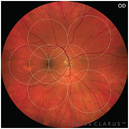
Virtually all commercially available imaging devices can capture posterior pole images. UWF images are available on a handful of devices, including the Heidelberg Spectralis, Natus RetCam3, Zeiss Clarus, and the Optos optomap systems. (Table.) There are no commercially available devices capable of panretinal imaging.
| Platform | Type of lens system | Single-capture field of view |
| Heidelberg Spectralis | Non-contact (optional, contact lens) | 55º (150º) |
| Natus RetCam3 | Contact | 130º |
| Zeiss Clarus 500 | Non-contact | 133º |
| Optos optomap | Non-contact | 200º |
Images from the different systems have slightly different appearances because they use different light sources, ranging from white light, red-green or red-green-blue laser wavelengths. All produce high resolution, finely detailed images.

All imaging devices can capture a wider field of view by having the patient look into various directions and then having software stitch these together into a single wider angle montaged image.
We chose the Optos optomap UWF platform, which uses confocal scanning laser technology and a specially designed ellipsoidal mirror capable of the largest available single-capture image (200°), even through an undilated pupil. Like most of the available systems, optomap is capable of a variety of imaging modalities including color, red-free, choroidal, fundus autofluorescence (FAF), fluorescein angiography (FA) and indocyanine green angiography (ICGA). The Optos platform offers a unique way of looking at the retina. The red and green channels may appear strange at first when compared with a “true color” white light UWF image. However, each of the two colors can be viewed separately, which gives us more information about the retina and choroid.
How do you capture an UWF image?
As just mentioned, the process for capturing an UWF image differs based on the platform you are using. Some, like ours, can produce an UWF image in a single capture. Others require montaging from two to seven smaller images.

Capturing an UWF optomap image is quite easy, and most of my staff learned how to operate the device in minutes. Patients sit directly in front of the device and look straight with their heads turned slightly to the side. The technician instructs them to align their eye with the highly visible blue internal fixation target, which changes to green when they are in the appropriate position (and red if they are too close). The patient’s eye is visible on the device tablet, and the technician raises or lowers the chinrest to help support the patient’s head at the correct height. With all the imaging devices, the manufacturers offer training on how to operate their device.
What are the benefits of UWF imaging?
First and foremost, UWF imaging makes me a better doctor and provides for better patient care. The ability to see into the far periphery facilitates the detection of pathology that might otherwise be missed, allowing me to diagnose and treat patients with greater accuracy and efficiency.
For diabetic patients, UWF is extremely valuable. The gold standard for evaluating and classifying DR has been the 7 standard photographic fields from the modified Airlie House classification from the Diabetic Retinopathy Study,2 subsequently modified by the Early Treatment Diabetic Retinopathy Study in the early 1990s.3 However, more recent research has shown that pathology frequently exists outside of this area. For example, UWF imaging often detects increased DR severity with higher risk of progression.4-6 UWF photography and fluorescein angiography have enhanced my ability to detect and assess peripheral disease including ischemia and neovascularization and has become an essential tool in assessing the baseline level of retinopathy at a patient’s first visit.
When a patient experiences flashers or floaters, in addition to scleral depression, UWF is always part of our exam, as it occasionally detects peripheral retina breaks missed by our examination.
In addition, ocular tumors would be nearly impossible to follow correctly without the use of UWF imaging, particularly those that are large or present in the periphery.
Along with assessing known diseases, UWF is a valuable tool for detecting and describing new pathologies as well as toxicities induced by certain systemic drugs.
UWF images are also a powerful patient education tool. This is particularly helpful with patients having difficulty understanding the need for follow-up appointments or treatment. For example, a diabetic patient may have normal vision despite potentially blinding peripheral proliferative disease. Reviewing their UWF images often makes the difference between a patient following through with our treatment plan or refusing treatment until it is too late.
An additional benefit we appreciate is the impact on practice flow. Single-capture UWF imaging is fast and efficient and has been shown to increase practice flow.7-9
Better serve patients
UWF is a unique technology that is easy to use and can help ophthalmologists and ophthalmic staff better serve their patients. OP
REFERENCES
- Choudhry N, Duker JS, Freund KB et al. Classification and guidelines for widefield imaging: recommendations from The International Widefield Imaging Study Group. Ophthalmol Retina 2019;3:843-849.
- Diabetic Retinopathy Study Research Group. Report 7. A modification of the Airlie House classification of diabetic retinopathy. Investigative Ophthalmology & Visual Science 1981;21:210-226.
- Early Treatment Diabetic Retinopathy Study Research Group. Grading diabetic retinopathy from stereoscopic color fundus photographs - an extension of the modified Airlie House classification: ETDRS report number 10. Ophthalmology 1991;98:786-806.
- Wessel MM, Aaker GD, Parlitsis G, et al. Ultra-wide-field angiography improves the detection and classification of diabetic retinopathy. Retina 2012; 32:785-791
- Silva PS, Cavallerano JD, Tolls D, et al. Potential efficiency benefits of nonmydriatic ultrawide field retinal imaging in an ocular telehealth diabetic retinopathy program. Diabetes Care 2014;37:50-55.
- Silva PS, Cavallerano JD, Sun JK, et al. Peripheral lesions identified by mydriatic ultrawide field imaging: distribution and potential impact on diabetic retinopathy severity. Ophthalmology 2013;120:2587-2595
- Lin C, Li AS, Ma H, et al. Successful interventions to improve efficiency and reduce patient visit duration in a retina practice. Retina March 18, 2021 (published ahead of print), doi: 10.1097/IAE.0000000000003169
- Nonmydriatic ultrawide field retinal imaging compared with dilated standard 7-field 35mm photography and retinal specialist examination for evaluation of diabetic retinopathy. Am J Ophthalmol. 2012 154;549-559.
- Real-Time Ultrawide Field Image Evaluation of Retinopathy in Diabetes Telemedicine program. Diabetes Care. 2015;38:1643-1649.


